


STAT: Quick MRI Could Improve Heart Failure Diagnosis, Guide Treatment
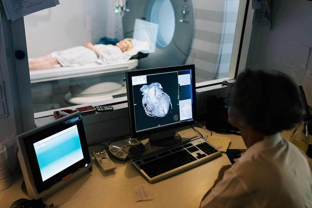
STAT recently interviewed Hsin-Jung Yang, PhD, director of Cardiac Imaging Research in the Biomedical Research Imaging Institute at Cedars-Sinai, about a promising new technique he helped develop that measures how much oxygen the heart uses during a routine cardiac MRI scan.
The experimental approach, described in a study published in Science Translational Medicine, could enable earlier and safer detection and monitoring of heart failure—a condition that affects 1 in 4 people in the U.S.
Yang, co-author of the study, told STAT the technique offers a quicker, less invasive way to assess cardiac oxygen consumption compared with current methods that require inserting catheters or radioactive tracers used in PET scans into patients.
He emphasized that measuring the heart’s ability to use oxygen efficiently reveals a key dimension of heart health—not only in heart failure, but in diabetes and hypertension as well.
“Those patients are more subject to cardiometabolic illness when the heart doesn’t function well and cannot pump as much blood as you need into your whole body,” Yang told STAT.
Yang and his team adapted a neuroimaging method that detects blood oxygen level-dependent signals in the brain and applied it to the heart.
The technique adds just three minutes to a standard cardiac MRI scan and does not require contrast agents, breath-holding or radiation exposure.
Because MRI is widely available in hospitals, Yang told STAT, the method could be used more broadly in lower-risk populations and repeated over time to monitor treatment response.
“People with obesity, diabetes, hypertension—those are key populations that we have the chance of doing earlier identification,” he said. “I think that is where the impact can translate the most.”
Read the complete article from STAT.