



Research Reveals Small Brain Area Has a Giant Role in Memory
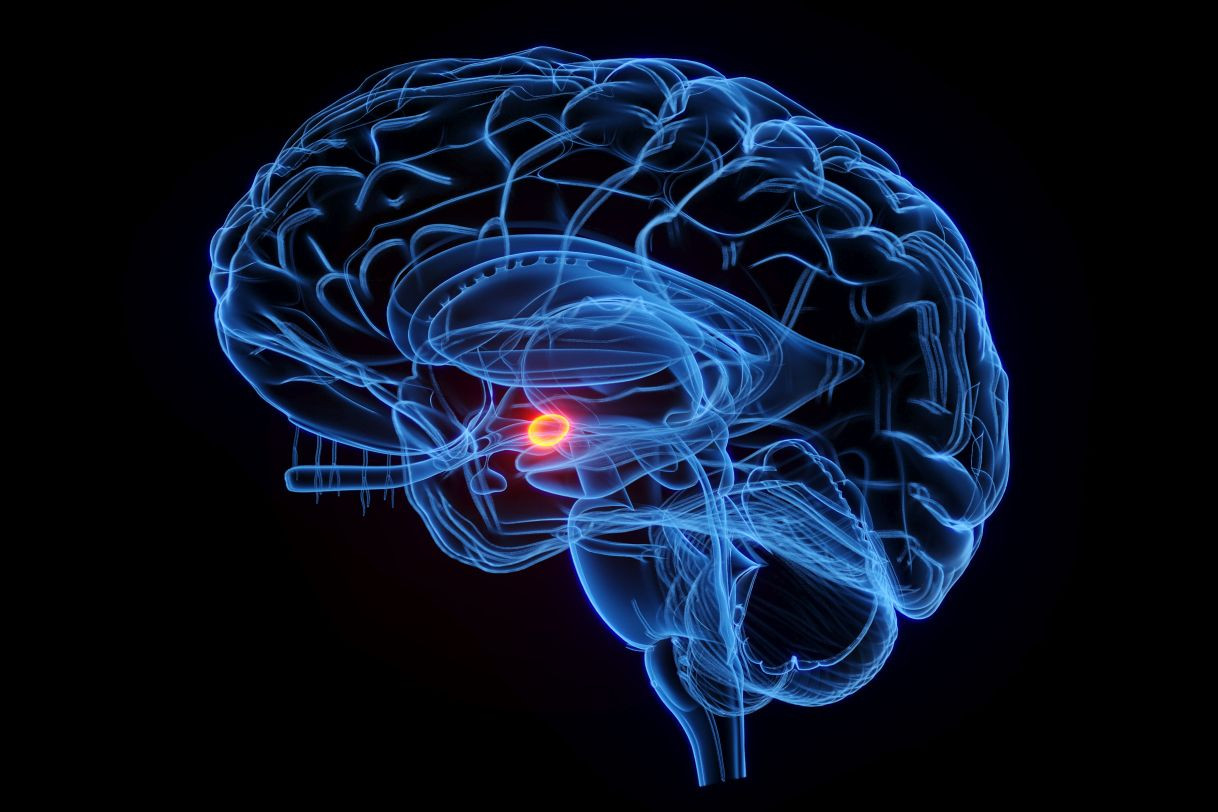
Walnut-Sized Amygdala Acts as Librarian to Categorize and Tag Images That Are Later Remembered, Cedars-Sinai Study Finds
A walnut-sized area of the brain known as the amygdala plays a giant role in determining which experiences end up in long-term memory and which are forgotten. The Cedars-Sinai Health Sciences University discovery points to the amygdala as a potential target for treating memory disorders.
 The amygdala, located deep below the brain surface in the temporal lobe, is classically associated with emotional and social behavior. In memory, its role was thought to be limited to learning and recalling experiences that invoked fear or other strong feelings. But the new study, published in the journal Nature Neuroscience, uncovered a much broader role for the amygdala in forming long-lasting memories in general.
In effect, the amygdala acts as a filter, tagging a small subset of ongoing experiences that end up being stored in long-term memory and thereby available for later retrieval.
“Our results demonstrate that the activity of specific kinds of neurons in the amygdala predicts, while the person sees an image for the first time, whether that image will later be remembered,” said Ueli Rutishauser, PhD, director of the Center for Neural Science and Medicine at Cedars-Sinai and the study’s corresponding author. “Such episodic autobiographical memories, such as where we parked our car or how we spent a recent family vacation, are critical to navigating everyday life and a sense of who we are.”
Investigators found that the brain’s ability to tag novel experiences for storage in long-term memory relies on specific types of neurons in the amygdala that respond selectively to specific categories of images, such as those showing faces, animals or landscapes. The stronger these category neurons reacted when a person viewed an image of the category to which the cell responded, the stronger that person's ability to later recall that image.
The study’s data comes from recordings of single-neuron activity in 112 surgical patients who had electrodes surgically implanted into their brains to monitor epileptic seizures. Participants were first presented with novel images they had never seen before and then asked to recall them later in the day.
 This finding is an example of the power of collaboration between scientists and physicians: It was the data collected across over 100 patients that allowed this key new scientific discovery, which was replicated in two different datasets in the study.
Collecting this large amount of data was made possible by a multi-institutional consortium led by Cedars-Sinai and funded by the National Institutes of Health (NIH) Brain Research Through Advancing Innovative Neurotechnologies (BRAIN) initiative.
“This is an impactful study that provides mechanistic insight into the role of the human amygdala—a region commonly associated with emotional responses—in determining which experiences and stimuli become lasting episodic memories. This rigorous human neuroscience study, which uses next-generation tools, has high potential to move discovery toward measurable health impacts,” said Mauricio Rangel-Gomez, PhD, program officer at NIH.
“Our findings have important clinical implications, as the memory impairments seen in disorders such as Alzheimer’s disease and temporal lobe epilepsy might be partly due to disrupted processes within the amygdala,” said Rutishauser, also a professor of Neurosurgery. “The amygdala might therefore be a suitable new target for treating these disorders using neuromodulation and/or cellular therapies.”
The research was led by Jiayang Xiao, PhD, who is the first author of the study.
Rutishauser added that future research could establish whether the amygdala plays a similar role in forming memories for other sensory modalities, such as sounds and smells.
“This study’s insights are the latest in a long line of research led by Cedars-Sinai and collaborators dedicated to revealing how human memory functions,” said Adam Mamelak, MD, director of the Epilepsy and Functional Neurosurgery Program and professor of Neurosurgery at Cedars-Sinai, and co-author of the study. “Beyond the pure scientific value of these breakthroughs, we are laser-focused on applying this new knowledge to improving the lives of patients.”
Additional Cedars-Sinai authors include Jonathan Daume, Natalia Kurilenko and Clayton Mosher.
Additional authors include Yousef Salimpour, William S. Anderson and Taufik A. Valiante.
Funding: This work was supported by the BRAIN Initiative through the National Institutes of Health (grant no. U01NS117839 to U.R.) and a Center for Neural Science and Medicine Postdoctoral Fellowship by Cedars-Sinai (to J.X.)
Cedars-Sinai Health Sciences University is advancing groundbreaking research and educating future leaders in medicine, biomedical sciences and allied health sciences. Learn more about the university.