


Research Tip Sheet: New Cedars-Sinai Findings in Cancer, Alzheimer's
The Latest Advances From Cedars-Sinai Investigators
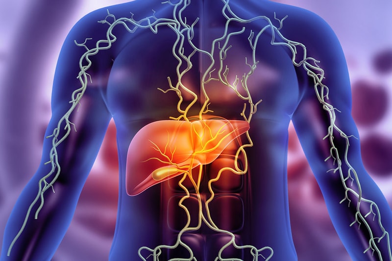 Immunotherapy Could Make Liver Transplants Possible for More Cancer Patients
Immunotherapy could make liver transplants an option for more patients with advanced liver cancer by shrinking their tumors, according to an international study co-led by Cedars-Sinai Health Sciences University investigators.
Liver transplants can cure liver cancer, but patients with large tumors and late-stage disease often don’t qualify. The study, published in Clinical Gastroenterology and Hepatology, found that when late-stage cancer patients received immunotherapy to shrink their tumors and then received transplants, they had outcomes similar to transplant patients whose cancer was diagnosed at an earlier stage.
“Our study provides important real-world evidence to guide the use of immunotherapy before liver transplantation in patients with liver cancer,” said Ju Dong Yang, MD, medical director of the Liver Cancer Program at Cedars-Sinai Cancer and senior and co-corresponding author of the study. “The findings support individualized treatment decisions and may help clinicians better identify suitable transplant candidates and optimize their outcomes.”
Investigators examined data from 408 patients who received immunotherapy before liver transplantation at 60 centers in 14 countries. They found that patients whose tumors were successfully reduced before transplantation had survival and cancer recurrence outcomes similar to those who met the transplant criteria before treatment.
Patients with a 90-day or greater gap between immunotherapy and transplantation had a lower risk of organ rejection. Tumor number and size, and the presence of a blood biomarker for liver cancer at transplantation, helped predict cancer recurrence.
“The findings may help clinicians assess patients’ risk of rejection and cancer recurrence, and optimize the timing between immunotherapy and transplantation,” Yang said, adding future prospective studies are needed to establish safer treatment strategies and improve long-term survival for liver cancer patients.
Additional Cedars-Sinai authors include Yee Hui Yeo, MD; Hasmik Adetyan, BS; Michael Luu, MPH; Irene Kim, MD; Alexander Kuo, MD; and Pin-Jung Chen, MD.
Other authors are listed in the manuscript.
Funding: This project did not receive any core funding; however, several authors have received support for their research, and this information can be found in the manuscript.
Disclosures: Dr. Ju Dong Yang reports the following conflicts of interest: consulting service for AstraZeneca, Eisai, Exact Sciences, and Fujifilm Medical Sciences.
 New Tool Could Predict Prostate Cancer Spread
A team led by Cedars-Sinai Health Sciences University investigators has created a tool that could predict whether prostate cancer will spread to other parts of the body. The tool, described in the Journal of Translational Medicine, could help doctors identify the men at highest risk of metastasis, which accounts for most prostate cancer-related deaths.
“By predicting whether a given patient’s prostate cancer will spread, our tool could help their care team decide whether to recommend prostate removal surgery or surveillance,” said Mohamed Omar, MD, assistant professor of Computational Biomedicine and Urology at Cedars-Sinai and corresponding author of the study. “And for patients who have surgery, it could eventually guide decisions about radiation or hormone therapy afterward.”
The tool, Met-Score, analyzes 45 genes from cells within a tumor and the cells that surround it. Investigators applied Met-Score to data on patients followed for many years after prostate removal surgery. Met-Score identified which patients were most likely to develop metastasis and separated intermediate-grade tumors into distinct high- and low-risk groups.
The Met-Score signature was detectable in patients’ diagnostic needle biopsies, and it distinguished patients whose disease was confined to the prostate at diagnosis from those whose cancer had already spread.
Investigators said that studies using Met-Score to evaluate prostate cancer patients at the time of diagnosis, then following those patients to compare their outcomes to Met-Score predictions, is the next step toward widespread clinical adoption.
Additional Cedars-Sinai authors include Itzel Valencia, Priyanka Vasanthakumari, Maryam Ranjpour Aghmiouni, Bryan Magaña, Minhyung Kim, Stephen J. Freedland, and Sungyong You.
Other authors include Pier Vitale Nuzzo, Edoardo Francini, Francesco Ravera, Giuseppe Nicolò Fanelli, Sara Bleve, Cristian Scatena, and Luigi Marchionni.
Funding: This work was supported by the Department of Defense grant HT9425-24-1-0381 (to M.O).
 Synaptic Changes Visible in the Retina Track Alzheimer’s Disease Severity
A new study by Cedars-Sinai Health Sciences University investigators linked loss of nerve cell connections and reduced levels of a key protein in the retinas of people with Alzheimer’s-related dementia with brain changes and cognitive decline. Their findings, published in Advanced Science, mean the eye may offer a window into early Alzheimer’s disease progression.
“Loss of synapses, which nerve cells use to communicate, is the best predictor of cognitive decline in Alzheimer’s,” said Maya Koronyo-Hamaoui, PhD, professor of Neurosurgery, Neurology and Biomedical Sciences at Cedars-Sinai and the senior and corresponding author of the study. “Our study shows this loss, and depletion of the protective protein UCHL1, which helps nerve cells function, begin in the retina as mild cognitive impairment sets in or even before symptoms arise.”
Because the retina can be viewed noninvasively, these findings lay the groundwork for early tracking of the disease and point to new ways of protecting synapses.
Investigators analyzed retinal tissue from people with normal cognition, mild cognitive impairment and Alzheimer’s disease. They also conducted experiments using nerve cells in the brain from humans and laboratory mice.
Investigators found that in the retina, synaptic connections were lost and UCHL1 levels declined as Alzheimer’s disease severity increased. They also found that exposure to amyloid beta, a protein associated with Alzheimer’s disease, reduced UCHL1 levels and synaptic connections even before nerve cells showed signs of degeneration.
“These findings advance our understanding of the brain-retina connection in Alzheimer’s disease and identify promising targets for tracking progress and preventing Alzheimer’s-related damage,” Koronyo-Hamaoui said. “Our goal is to translate these insights into noninvasive tools for earlier detection and more precise monitoring of disease progression and treatment response.”
First author is Altan Rentsendorj. Additional Cedars-Sinai authors include Jean-Philippe Vit, Alexandre Hutton, Yosef Koronyo, Saba Shahin, Edward Robinson, Ousman Jallow, Julia Sheyn, Bhakta Prasad Gaire, Lalita Subedi, Miyah R. Davis, Barathan Gnanabharathi, Alexander V. Ljubimov, Katlin Silm, Keith L. Black, Jesse G. Meyer, and Dieu-Trang Fuchs.
Other authors include Alfredo A. Sadun, Ernesto Barron, Anthony Rodriguez, Stuart L. Graham, Vivek K. Gupta, Mehdi Mirzaei, Debra Hawes, and Lon S. Schneider.
Funding: This work was supported by the National Institutes of Health (NIH)/the National Institute on Aging (NIA) through the following grants: R01AG056478, R01AG055865, and AG056478-04S1 (MKH), the National Institute for General Medical Sciences: R35GM142502 (JGM), the Hertz Innovation Fund, and the Gordon, Goldrich, Wilstein, and Saban Private Foundations (MKH). M.R.D. and E.R. were supported by the Ray Charles Foundation.
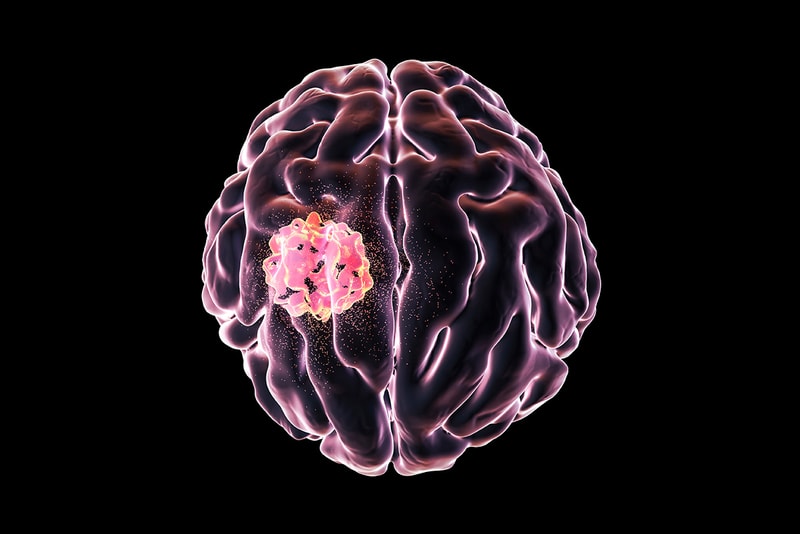 Researchers Develop More Accurate Model of Aggressive Brain Cancer
Cedars-Sinai Health Sciences University investigators helped create a new and improved scientific model of glioblastoma, the most common and aggressive brain cancer in adults. Their work, published in Cancer Research, could help scientists better understand the disease and evaluate new therapies.
“Past studies using the few existing glioblastoma models have indicated that treatments such as immunotherapy would be effective in patients, but that turned out to be inaccurate,” said Stephen Shiao, MD, PhD, professor of Radiation Oncology and Biomedical Sciences at Cedars-Sinai and co-corresponding author of the study. “Our model more accurately reflects patient tumors, and we believe it will better predict the effectiveness of treatments.”
Shiao said glioblastoma treatment options have changed little in decades, and the five-year survival rate overall remains below 7%.
Investigators used a technique called mosaic analysis by dual recombinase-mediated cassette exchange (MADR) to create tumors in laboratory mice. The tumors carried genetic mutations commonly found in glioblastoma tumors in human patients, including changes that lock cell growth into the “on” position.
The study found that tumors created using MADR exhibited the same characteristics as tumors in human patients. Cancer cells from the tumors migrated outward into healthy brain tissue, the environment surrounding the tumors had few cancer-killing T-cells, and the tumors were resistant to immunotherapy treatments.
Investigators also found that the tumor cells were better than those in previous preclinical models at evading the immune system, similar to human tumor cells.
Katie Grausam, PhD, who was a project scientist in the Shiao Lab and co-first and corresponding author of the study, said this modeling system could be used to build “avatar” models based on different tumor mutations, allowing for personalized treatment and evaluation of treatment response.
Additional Cedars-Sinai authors include David Rincon Fernandez Pacheco, Emily A. Hatanaka, Eddie Lu, Paul W. Linesch, Naomi Kobritz, Xuemo Fan, and Moise Danielpour.
Other authors include Marissa S. Pioso, Julio C. Sanchez, Marissa Li, Gilbert Herrera, Thomas J. Lai, Trinh L. Phan, Sophie Ashley, Ariel A. Andrade, Lauren Markus, Jingwen Yao, Bohyeon Yu, Aaron A. Diaz, Linda M. Liau, David A. Nathanson, Robert M. Prins, and Joshua J. Breunig.
Funding: This study was funded in part by the National Institutes of Health, National Cancer Institute SPORE in Brain Cancer (grant no. 2P50CA211015 to D.A.N. and R.M.P.), the IMAT Supplement (grant no. P50CA211015-07S1 to D.A.N. and R.M.P), and grant no. R33CA278564 (to J.J.B) , R33CA236687 (to J.J.B), R01NS131782 (to J.J.B.) and P50CA211015-05S1 (to J.J.B.), and the CureSearch Acceleration Initiative Award (to J.J.B.), and the Department of Defense (award no. NF220040 and KC200178 to J.J.B.). Additional support was provided by NIH Ruth L. Kirschstein Institutional National Research Service Awards (grant no. T32CA009056 to M.S.P., grant no. T32CA009120 to J.C.S.), the NIH Ruth L. Kirschstein National Research Service Award Individual 20 Predoctoral Fellowship (grant no. F31CA294887 to M.S.P.), the UCLA Chancellor’s Postdoctoral Fellowship Program (to J.C.S.), the 2022 Cedars-Sinai Cancer Development Funds for Team-Based Science Award (to J.J.B.), and the Parker Institute for Cancer Immunotherapy. Research reported here included work performed in the Technology Center for Genomics and Bioinformatics (TCGB) and JCCC Flow Cytometry Core supported by NCI/NIH awards P30CA016042 and 5P30AI028697.
Cedars-Sinai Health Sciences University is advancing groundbreaking research and educating future leaders in medicine, biomedical sciences and allied health sciences. Learn more about the university.