


A Bigger, Better AI Tool for Interpreting Common Heart Test
Study Co-Led by Cedars-Sinai Produces Largest-Ever Model for Analyzing Echocardiography Reports
Investigators from Cedars-Sinai and other institutions have created the largest artificial intelligence-based model for analyzing and producing reports from echocardiograms, ultrasound images doctors often use to diagnose heart disease. Their study, published in Nature, marks a major step toward bringing automated echocardiography reports into patient care.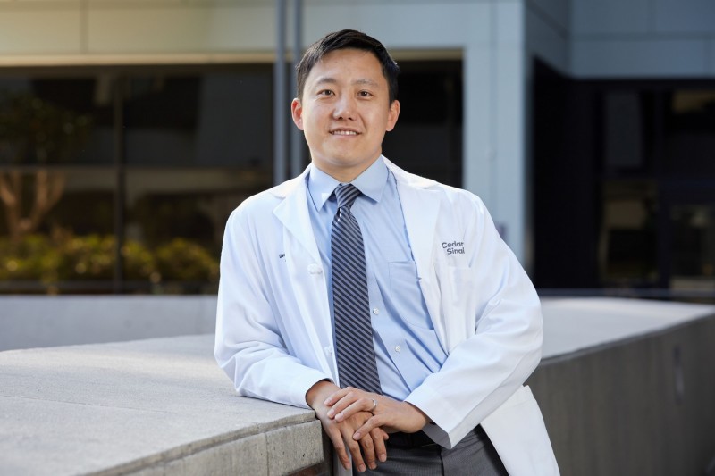
An echocardiogram uses ultrasound to create images of the heart and the results are analyzed by cardiologists or sonographers trained to read the results.
“Our AI model addresses a critical need,” said David Ouyang, MD, adjunct assistant professor in the Smidt Heart Institute at Cedars-Sinai and co-corresponding author of the study. “Many hospitals and clinics, especially in rural areas, do not have cardiologists or sonographers on staff. By generating accurate automated reports for physicians, we could improve access to this important diagnostic technology.”
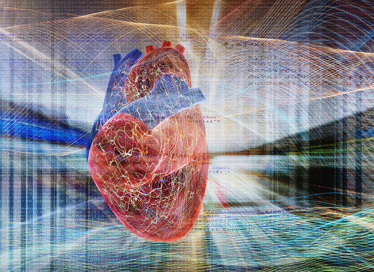
The AI model investigators created, called EchoPrime, is a type known as a foundation model. These models are trained on massive datasets and can perform multiple tasks, including assessing ultrasound images and evaluating clinical report text. EchoPrime was trained on greater than 10 times more data than prior AI foundation models for echocardiography, making it the largest such model to date, according to the investigators. EchoPrime analyzes ultrasound images, then produces a verbal summary of heart function and structure meant to assist clinicians in diagnosing a patient.
The team trained EchoPrime on more than 12 million videos paired with cardiologists’ written interpretations of the tests. They then tested the model on data from Kaiser Permanente Northern California, Cedars-Sinai Medical Center, Stanford Health Care, Beth Israel Deaconess Medical Center in Boston, and Chang Gung Memorial Hospital in Taiwan. The investigators 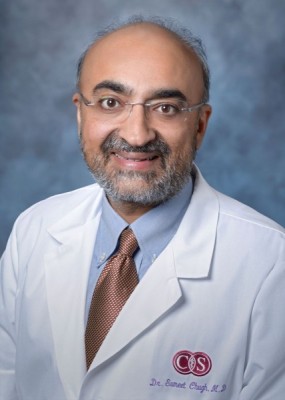 found that EchoPrime outperformed other foundation models for interpreting echocardiograms and matched or exceeded the performance of AI models designed to perform a single ultrasound task. It was also able to detect rare diseases of the heart.
The investigators are now conducting randomized clinical trials that compare reports generated by EchoPrime with those produced by cardiologists and sonographers. The goal is to confirm the accuracy of the model and ensure it is useful to cardiologists.
“This remarkable study from Dr. Ouyang and colleagues builds on several years of AI-enhanced imaging science at Cedars-Sinai,” said Sumeet Chugh, MD, vice dean and chief artificial intelligence health research officer at Cedars-Sinai. “EchoPrime is a unique foundational model that could potentially democratize AI-powered echocardiography interpretation on a global scale.”
Other Cedars-Sinai authors include Milos Vukadinovic, I-Min Chiu, Debiao Li and Susan Cheng.
Other authors include Xiu Tang, Neal Yuan, Tien-Yu Chen, Paul Cheng and Bryan He.
Funding: D.O. discloses National Institutes of Health NHLBI grants R00HL157421, R01HL173526 and R01HL173487.
Cedars-Sinai Health Sciences University is advancing groundbreaking research and educating future leaders in medicine, biomedical sciences and allied health sciences. Learn more about the university.