


Discoveries
Stop Signal Neurons
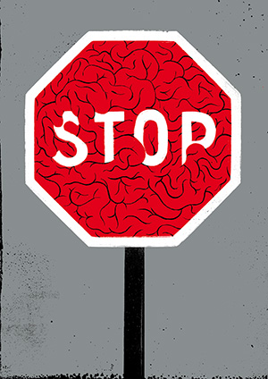
We've all done it—taken our foot off the brake while stopped at a traffic light, only to reapply it because the signal is still red. That immediate correction is triggered by the appropriately named "stop signal neurons," charged by our brain with canceling potentially harmful actions. When this automatic ability fades, it can lead to the unwanted movements associated with Parkinson's disease and other neurological disorders.
Parkinson's disease, which affects nearly 1 million people in the U.S. alone, leads to a paradoxical mixture of uncontrolled, involuntary movements and an inability to move. How this process occurs and what regions of the brain are responsible for it have remained elusive despite years of intensive research.
Now, Cedars-Sinai investigators have solved part of this longstanding mystery. "This first-in-human study identifies the underlying brain processes required for stopping movements," notes Ueli Rutishauser, PhD, the Board of Governors Chair in Neurosciences.

Investigators studied patients with Parkinson's disease undergoing deep-brain stimulation, a common surgery for the disease that implants tiny electrodes in the basal ganglia—the brain region that regulates motor control—to block excessive movement.
The researchers discovered that neurons in one part of the basal ganglia region—the subthalamic nucleus—indicated the need to "stop" an already initiated action. These neurons responded very quickly after the appearance of the stop signal. They had never before been observed "in action" in humans.
"This discovery provides the ability to more accurately target deep-brain stimulation electrodes," says Adam Mamelak, MD, co-director of the Pituitary Center at Cedars-Sinai and director of the Functional Neurosurgery Program.
