Back on Her Feet Thanks to CSF Leak Repair
Date
November 1, 2017

Date
November 1, 2017
Credits
Medical providers featured in this article
In Brief
{{cta-block}}
After Excruciating Headaches Kept Her Bedbound For Weeks, New Mexico Woman Credits Cedars-Sinai With Recovery.

When Beth Johnson stood up from working at her computer one beautiful October evening, she noticed "a kind of funny feeling" in the back of her neck. Five days later, the sensation had become an excruciating headache that came on whenever she tried to stand or sit upright.
Her symptoms and magnetic resonance imaging (MRI) results suggested that a hole had developed in the lining around her spinal cord, known as the dura. The resulting loss of cerebrospinal fluid (CSF), which surrounds and protects the brain and spine, caused Johnson’s brain to shift and sag away from her skull when she tried to stand. This led to severe positional headaches and eventually periods of confusion.
"I have said to Dr. Schievink repeatedly, 'I couldn't stand. I couldn't sit. And my brain was being totally compromised. You saved my life. I am so grateful."

After two attempts at another neurosurgical center failed to patch the hole, Johnson, who lives 13 miles from Santa Fe, N.M., came to Cedars-Sinai, where Wouter Schievink, MD, director of microvascular neurosurgery and the CSF Leak Program in the Department of Neurosurgery, surgically repaired the tear.
Schievink, one of the world's most experienced CSF leak specialists, said Johnson's tear was extensive but no larger than many he has treated. It was, however, located on the front side of her spinal cord, making surgical access more challenging and the repair procedure more delicate.

When Johnson, a self-described fast-moving, quick-thinking, Type-A personality, initially felt the strange sensation in her neck, she didn't slow down at first. But as symptoms got worse, she wondered if she should go to an urgent care center. Friends suggested she was simply “stressed out” and recommended therapeutic massage. When she was no longer able to sit or stand without pain, however, she broke down and saw her primary care physician. Her doctor immediately sent her for MRIs that showed she had spontaneous intracranial hypotension. A defect in the dura had opened, allowing the CSF to leak.
Two days later, Johnson was flown by air ambulance to another neurosurgical center, where doctors used injections of her own blood to try to seal the tear. This treatment, known as an epidural blood patch, often is used to treat CSF leaks that will not heal on their own. Johnson was allowed to go home a week later, but soon it was clear to her and Char Schnepf, her partner of 15 years, that the treatment had not worked for her.
"When I called the doctors, they said to come back and have a third blood patch. They said sometimes people need 10 of them,” said Johnson, manager of a family compound and a life transitions coach. “This didn't make sense to me."

At the suggestion of a client and friend who had found Schievink's name online, Johnson called Cedars-Sinai. Schnepf wrote a letter, collected copies of the original MRIs and shipped them overnight to Schievink.
"He looked at my MRIs and called me the next day," recalled Johnson, who again was flown by air ambulance, this time to Los Angeles. She met Schievink the afternoon she arrived and immediately underwent a series of diagnostic procedures. They confirmed the defect's location at the front of the spine and showed a calcium deposit on a disc, which may have contributed to the tear.
But working from the back to shave down the calcification and repair the lining at the front of the spinal canal is not straightforward. The spine's large, drum-shaped bones, called vertebral bodies, are stacked on the front side of the spinal canal, and separated by discs that act as cushions. Each vertebral body has a hard, bony structure called a pedicle that consists of two parts – left and right. These form the sides of the spinal canal and connect the large bones to the smaller ones in back. The entire structure protects the spinal cord, nerves and nerve roots that carry electrical signals to and from the brain.
"We removed a piece of bone from the back part of the spinal canal and took out half of one of the pedicles. By removing half of a pedicle, I could rotate the spinal cord away from where the tear was, which gave me enough room to work. I closed the tear with four sutures, which I'm confident will provide a strong and permanent repair," Schievink said.
"We always use advanced technology to frequently monitor the spinal cord – before and after each suture is placed," he added. "When her last suture was in place, the monitor detected a reduced signal to her right leg, and she woke up with some weakness in that leg, which resolved overnight. But she had considerable weakness resulting from weeks spent incapacitated. That kind of generalized deconditioning doesn't improve immediately, and she would need a course of physical therapy to really get back on her feet."
In Discoveries: Better Brain Surgery
After surgery, Johnson remained quiet and flat in bed for a day. She sat up in a chair the following Sunday afternoon. It was the first time in more than a month that she had been upright for any real length of time. She was able to walk with a walker the next day.
An MRI done three days post-surgery confirmed what already was obvious to Johnson: The leak was closed and the CSF was steadily returning to its normal level.
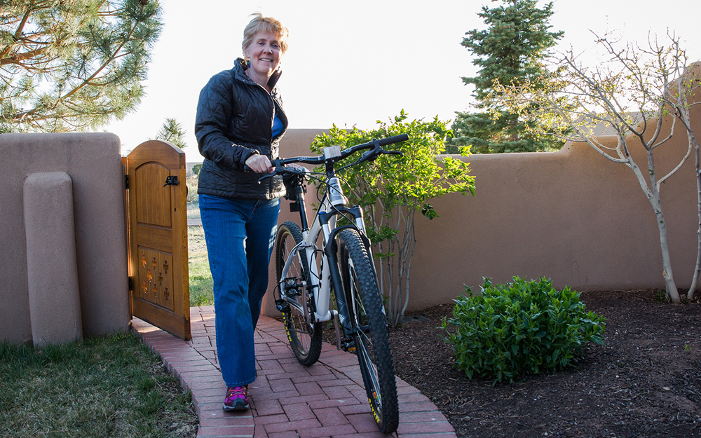
Johnson likes to ride her bike and participate in yoga regularly. She is working hard to rebuild the muscle mass she lost, and is regaining the mental quickness she had before her ordeal.
"I have said to Dr. Schievink repeatedly, 'I couldn't stand. I couldn't sit. And my brain was being totally compromised," Johnson said. "You saved my life. I am so grateful."