Research Areas
Microgel-encapsulated stem cells to alleviate low back pain
Chronic low back pain affects millions of people worldwide and is closely associated with intervertebral disc (IVD) degeneration. Current treatment methods such as surgical intervention and pain management focus on alleviating symptoms of the disease without addressing the disease itself. Our lab is looking to develop a stem cell therapy to regenerate the degenerated IVD by leveraging microgel technology to safely deliver induced notochordal cells into the disc. By encapsulating cells within microgel we can inject the cells directly into the IVD without the need for surgery. The microgel also has added benefits of protecting the cells from the harsh environment of the degenerated IVD, the possibility of pre-conditioning the cells in vitro, and shielding the cells from shear force exerted by pushing them through a syringe and needle. Research will be done in several pre-clinical models to determine the efficacy and safety of the microgel treatment. This study is funded by the California Institute for Regenerative Medicine (CIRM) and the NIH HEAL Initiative.
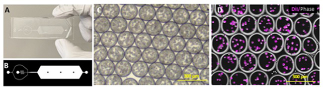
Microgel encapsulation of nucleus pulposus cells. A-B: Sample layout of the microfluidic device used to encapsulate microgels. C: Phase image of cell loaded microgels in microfluidic device. D: Fluorescence image of DiD labeled cells in microgels.
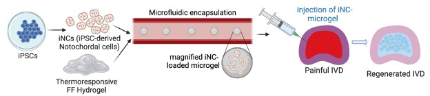
Cartoon layout of the injection of microgel encapsulated stem cells into a degenerated IVD.
Studying the mechanisms of discogenic low back pain
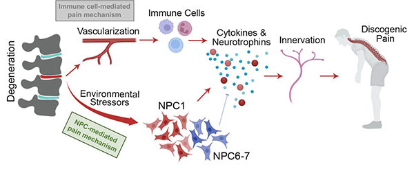
Low back pain is the leading cause of disability, primarily caused by degenerating intervertebral discs of aging individuals. Low back pain is a common problem that affects many people, and a significant portion of it is caused by the deterioration of discs in the spine. However, we don't fully understand why some deteriorating discs cause pain while others don't. We believe that certain environmental factors affect specific cells in the discs, which then lead to the onset of low back pain. To investigate this, we compared samples of degenerated disc tissue from surgical discards and human cadavers. We used a technique called single-cell RNA sequencing to analyze the cells in these discs. We looked at three discs that were associated with back pain and four discs that did not cause any symptoms. We specifically focused on a type of cells called nucleus pulposus cells (NPCs) from these discs. We then took NPCs from the discs that caused back pain and NPCs from the pain-free discs for further analysis. We cultured the NPCs in a lab and exposed them to different types of stress. We wanted to see if the stressed NPCs would have any pain-inducing effects. We conducted experiments both in the lab using special devices and in rats. By analyzing the data, we found that a particular group of NPCs associated with the discs that caused back pain. In the animal study, we injected the stressed NPCs into the discs of rats, which resulted in the expression of pain-related markers and increased pain behaviors in the rats compared to the control groups.
This study reveals that a specific type of stressed NPCs is responsible for triggering low back pain. Understanding this mechanism could help develop targeted therapies for low back pain that focus on these specific cells, instead of regenerating the entire discs.
Developing stem cell therapies for tendon injuries
Tendon injury is one of the most prevalent musculoskeletal problems in the U.S. affecting an estimated 33 million people every year. An injured tendon rarely achieves the level of functionality it had prior to injury, resulting in long rehabilitation periods and limited mobility. Development of a cell and tissue engineering approach for repairing tendons has the potential to dramatically improve patient outcomes. In the Sheyn Lab, we are focused on two main approaches: 1) elucidating the pathways involved with tendon cell fate by capitalizing on induced pluripotent stem cell (iPSC) and single-cell RNA sequencing technologies, and 2) implementing this knowledge in combination with tissue engineering techniques and biomaterials to develop more effective alternatives for traditional tendon repair using animal models. The first step to developing a therapeutic solution for tendon repair is to better understand how tendons develop and differentiate from their origins. To do so, we are investigating the signaling pathways involved with tendon differentiation and maturation. Informed by single-cell RNA-sequencing technology, we can differentiate human iPSCs to tenocyte precursor cells in a stepwise manner using chemically defined media and small molecules. Optimization and further characterization of these cells will not only allow us to better understand the mechanisms behind tendon development, but also allow us to look into applying these cells for in vivo applications. This study is currently funded by the CIRM.
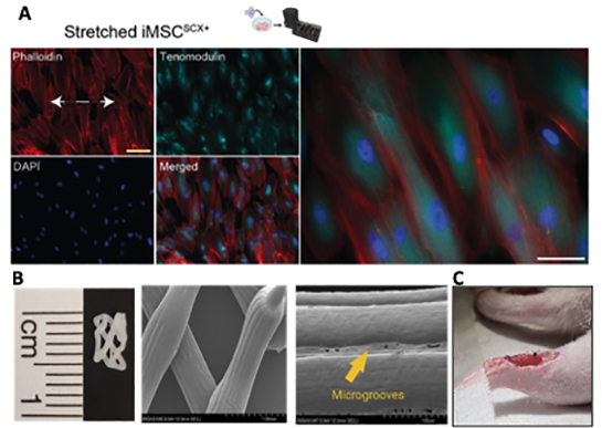
A: Immunocytochemistry cytoskeleton visualization Phalloidin and expression of tenogenic marker TNMD demonstrate tenogenic differentiation of iTenocytes after cyclic loading in a 2D bioreactor. (Papalamprou et al) B: Custom polycaprolactone scaffold with microgrooves micro-topography imaged via scanning electron microscope (Kaneda et al). C: iTenocyte-seeded collagen scaffold sutured into a injured rat Achilles tendon.
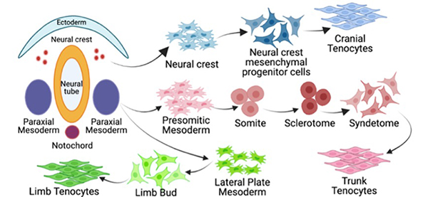
Tenogenic pathways in embryogenesis.
Injectable human iPSC-derived macrophages as a potential preventive treatment for knee post- traumatic osteoarthritis (PTOA) in a rat model
The dysregulated balance between tissue-resident and bone marrow-derived macrophages (MACs) has been suggested to be critical in the development of knee osteoarthritis (OA), a degenerative cartilage disease that is often associated with pain. Normalizing the aberrant M1/M2 ratio of MACs has been suggested as a therapeutic strategy for macrophage-involved diseases. An innovative source for the standardized in vitro generation of MACs is induced pluripotent stem cells (iPSCs), since iPSCs provide an unlimited pool of polarized MACs without donor age restrictions.
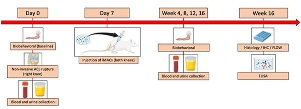
This study section focuses on the hypothesis that polarized iMAC therapy may modify the pro-inflammatory environment of ACL-torn knees, and consequently hinder the onset of knee progression and associated pain through normalization of an aberrant M1/M2 ratio in a rat PTOA model. Using this model, the efficacy of iMAC therapy in prevention of PTOA onset and progression will be tested. Induced MAC and placebo (saline) injection into the injured knee will be performed and followed for 16 weeks. Outcome measures include cartilage quality and thickness as well as inflammatory and pain responses.
Contact the Sheyn Lab
127 S. San Vicente Blvd.
Advanced Health Sciences Pavilion, A8308
Los Angeles, CA 90048