


Research Tip Sheet: Immunity, Cancer Wasting, Eye Disease
The Latest Advances From Cedars-Sinai Investigators
 Revealing How Transplanted Neural Stem Cells Preserve Vision
Cedars-Sinai investigators working to optimize a cell-based treatment for retinitis pigmentosa have uncovered how transplanted neural stem cells interact with host retinal cells to preserve vision. The findings, published in Nature Communications, may guide future research toward strategies to treat degenerative eye disease.
“We used single-cell analysis to show that neural stem cells can protect vision in several ways, including providing protective proteins, restoring retinal cells to a healthier state, reducing cellular stress, and maintaining retinal integrity,” said Clive Svendsen, PhD, executive director of the Board of Governors Regenerative Medicine Institute and co-corresponding author of the study.
Investigators transplanted neural stem cells into the retinas—the light-sensitive tissue lining the back of the eye—of laboratory rats with retinal degeneration. Previous studies have shown the transplants significantly reduced vision loss in the animals for up to 180 days, the equivalent of about 20 years in humans. In this study the team examined interactions between the transplanted cells and diseased retinal cells to better understand the neural stem cells’ protective effects.
“Our study reveals that the interaction between neural stem cells and host retinal cells dynamically changes over time,” said Shaomei Wang, MD, PhD, professor of Biomedical Sciences and co-corresponding author of the study. “Through a better understanding of this process, we may be able to develop more powerful approaches to treat eye diseases in the future.”
Investigators are now evaluating the use of neural stem cells engineered to express key protective proteins identified in this study to further improve the host retinal environment.
Additional Cedars-Sinai authors include Saba Shahin, Shaughn Bell, Bin Lu, Hui Xu, Jason Chetsawang, Stephany Ramirez, Jorge S. Alfaro, Alexander Laperle and Soshana Svendsen.
Other authors include Somanshu Banerjee and Vivek Swarup.
Funding: This work was supported by the California Institute Regenerative Medicine (LSP1-08235). J.C. was supported by CIRM-EDUC-08383 and S.R. was supported by CIRM-EDUC2-12638, and funding from the Board of Governors Regenerative Medicine Institute at Cedars-Sinai Medical Center.
 Preclinical Study: Protein Regulates Autoimmune Response
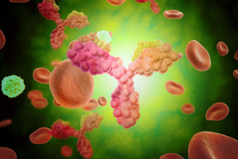
Cedars-Sinai Health Sciences University investigators have identified for the first time a protein’s role as a “dimmer switch” that can calm an overactive immune system and restrain harmful inflammation. The protein, Butyrophilin 2A2 (BTN2A2), interacts with a key molecule that controls the strength of T-cell responses.
The findings, published in Nature Communications, define a unique pathway that helps balance immune activity and could be harnessed to limit damage caused by a variety of autoimmune diseases.
In laboratory mice, loss of BTN2A2 led to exaggerated immune reactions and an increase in damaging kidney inflammation called glomerulonephritis. Treatment with BTN2A2 reduced disease severity by increasing immune-regulating T cells and lowering inflammation.
Supporting laboratory experiments in human T-cells demonstrated similar immune-calming effects.
“Glomerulonephritis remains a leading cause of chronic kidney disease and kidney failure worldwide, with limited treatment options,” said Ananth Karumanchi, MD, co-corresponding author of the study and director of the Renovascular Research Center at Cedars-Sinai. “Our findings provide a strong foundation for future studies aimed at modifying immune-driven kidney disease rather than simply managing its symptoms. The pathway could also be targeted in a range of autoimmune and inflammatory diseases including rheumatoid arthritis, multiple sclerosis, inflammatory bowel disease, and transplant rejections.”
Other Cedars-Sinai authors include Shafat Ali, Anders H. Berg, Michifumi Yamashita, Ambart E. Covarrubias, Jordan Mundell, Pranali N. Shah, Ruan Zhang, Vincent Dupont, Bong-Ha Shin, Shen Yang, Madhusudhanarao Katiki, Ramachandran Murali, Margareta D. Pisarska, Ravi Thadhani, Peter S. Heeger and Stanley C. Jordan
 Pinpointing Indicators of Cancer Wasting Syndrome
An analysis of biomarkers in patient blood samples by Cedars-Sinai Health Sciences University investigators could help with early detection of cachexia, or cancer wasting syndrome.
The study, published in Cancers, explores biologic signals detectable in the blood that could be used to design future strategies for assessing patient risk and develop therapies aimed at mitigating fatigue and muscle and fat loss experienced by many patients with cancer.
“We found that in patients with advanced non-small cell lung cancer, cachexia biomarkers change over time,” said Kamya Sankar, MD, co-medical director of the Thoracic Disease Research Group at Cedars-Sinai Cancer and corresponding author of the study. “And treatments targeting one of the early cachexia biomarkers we identified, an inflammatory protein called GDF-15, are already under evaluation in clinical trials.”
Investigators measured the blood of 27 patients with non-small cell lung cancer at two different time points. In patients with early cachexia, they found higher levels of inflammatory proteins such as GDF-15. In patients with later-stage cachexia, they found increased mitochondrial DNA, which comes from the parts of cells that convert food into energy.
Larger, prospective studies are required to validate the clinical benefit of these biomarkers, but they could serve as the basis for risk assessment of patients and may inform design of future clinical trials of therapies for cancer-associated cachexia, Sankar said.
Additional Cedars-Sinai authors include Elham Kazemian, Nicole Lorona, Carlos D. Cruz-Hernández, Mitra Mastali, Akil A. Merchant, Jennifer Van Eyk, Karen L. Reckamp, Neil A. Bhowmick, and Jane C. Figueiredo.
Other authors include Alex K. Bryant and Puneeth Iyengar.
Funding: This work was supported by the U.S. National Cancer Institute (U54CA260591, PI Figueiredo), Department of Defense (LC240075, PI Sankar), and Cedars-Sinai CTSI grant (UL1TR001881, PI Sankar).
Cedars-Sinai Health Sciences University is advancing groundbreaking research and educating future leaders in medicine, biomedical sciences and allied health sciences. Learn more about the university.