


A Telegram From Your Cells

Cedars-Sinai Cancer Investigators Use AI Technology to “Fingerprint” Messages Sent Between Cells, Paving the Way for a New Understanding of Health and Disease
The body’s cells communicate with each other via biological “telegrams” called extracellular vesicles (EVs). Cedars-Sinai Cancer investigators have created the first system for profiling EVs so that scientists can begin to understand their messages. Their study, published in the peer-reviewed journal ACS Nano, is the first step toward a better understanding of EV biology and the development of new clinical tests to measure disease progression in real time.
“Our hope is to be able to associate different EV fingerprints with different diseases and responses to treatment,” said Dolores Di Vizio, MD, PhD, professor of Urology, co-leader of the Cancer Biology Program at Cedars-Sinai Cancer and co-senior author of the study. “As cancer scientists, we are primarily interested in applying this new technology to the creation of ‘liquid biopsy’ tests for cancer detection. However, because all cells in the body produce EVs, we also envision its future use to identify biomarkers of cardiovascular disease, autoimmune diseases or neurological disorders.”
EVs are made of an outer envelope of fat molecules filled with “cargo” that might include RNA, proteins and lipids. The tiny particles vary greatly in size, with the largest being about one-tenth the width of a human hair.
“Prior to this study, EVs were mainly classified by their size and by how they were generated,” said Andries Zijlstra, PhD, adjunct associate professor of Pathology, Microbiology and Immunology at Vanderbilt University Medical Center and co-senior author of the study. “The goal of this study was to create a technology that could classify EVs based on several of their characteristics and their various combinations.”
EVs are studied using flow cytometry. The particles are separated from a blood sample, then suspended in fluid flowing through a laser beam that measures their physical and chemical characteristics.
To create the new technique of EV fingerprinting, investigators measured 20 different EV characteristics and used machine learning to classify various combinations of membrane composition, cargo, size and method of generation.
“The concept is that cells in different disease states will produce EVs with a unique fingerprint,” Di Vizio said. “For instance, we have prostate cancer cell lines, and each of these lines now has an EV fingerprint associated with it.”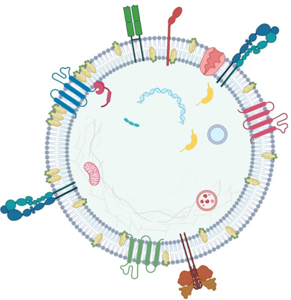
A further advantage of EV fingerprinting is that it can be performed directly from a blood sample without the laborious process of separating out the EVs, Di Vizio said. This means the technology can be used to develop “liquid biopsy” tests that can be used in clinics.
“Our EV fingerprinting method is faster than most liquid biopsy approaches that require the isolation of EVs,” Di Vizio said. “These tests could be used for diagnosis, monitoring progression of disease and response to treatment, and perhaps even predicting patient outcomes.”
Investigators will next explore pairing specific EV fingerprints with specific disease states, beginning with aggressive forms of cancer, Di Vizio said.
“This type of translational science is key to our mission at Cedars-Sinai Cancer,” said Dan Theodorescu, MD, PhD, director of Cedars-Sinai Cancer and the PHASE ONE Foundation Distinguished Chair and Director at the Samuel Oschin Comprehensive Cancer Institute. “By developing practical new testing techniques that allow us to precisely measure the state of a patient’s health, we can promote the concept of ‘precision prevention,’ expand the promise of precision therapy in oncology and save patients’ lives.”
Funding: This work was supported in part by grants from the National Institutes of Health (R01CA218526 to AZ and DDV; R01CA234557 to 1183 DDV; R01CA249424 to HP and AW; P01CA229123, R01CA206458, R01CA249684 and U01CA224276 to AMW) and the National Science Foundation (NSF-2328276 and NSF-2036809 to AMW and JTW).
Additional authors include Tatyana Vagner, Ariana K. von Lersner, Fabiane Fernandes, Patricia Midori Murobushi Ozawa, Marques Jackson, Matthieu Masureel, Hoangdung Ho, Sierra M. Lima, Tatyana Vagner, Bong Hwan Sung, Mohamed Wehbe, Kai Franze, Heather Pua, John T. Wilson, Jonathan M. Irish and Alissa M. Weaver.
Read more on the Cedars-Sinai Blog: Study Sheds Light on How Cancer Spreads in Blood