Why Breast Cancer Screening Should Be Individualized
Date
October 15, 2026
Credits

Date
October 15, 2026
Credits
Medical providers featured in this article
In Brief
Most women assume breast cancer screening is straightforward: You reach a certain birthday, schedule a mammogram and repeat at regular intervals. Screening recommendations are all over the map, though. Trusted medical organizations are split on when breast cancer screening should begin, how often it should occur, and which supplemental imaging is appropriate—and when.
“Instead of treating these guidelines as strict rules that you have to decode alone, view them as an invitation to start a conversation with your physician,” said Carey Cullinane, MD, a breast surgical oncologist at Cedars-Sinai.
The best breast cancer screening is not a single test performed annually after you reach a certain age. It’s an ongoing dialogue with your physician about creating a screening schedule that makes sense for your individual risk—and that discussion should begin well before you turn 40.
{{providers}}
Decoding Breast Cancer Risk
Understanding your personal breast cancer risk is more important than ever. The average woman in the U.S. has 13% chance of developing breast cancer in her lifetime, up from less than 10% in the 1970s. Risk is rising fastest among women once considered “low risk”: those under age 50, and sometimes even under 40.
Most screening guidelines are built for women at “average risk.” If you’re at higher risk, your recommendations may look different. The trouble is, most women don’t know which category they fall into.
“Before deciding on a screening schedule, women should sit down with their physicians—ideally in their 20s or 30s—to better understand their personal risk for developing breast cancer,” said Maxine Jochelson, MD, director of mammography for Breastlink in Beverly Hills/RadNet, a Cedars-Sinai partner organization.
Free online risk calculators, such as the Gail Model and the Tyrer-Cuzick Risk Assessment Calculator incorporate multiple factors, including age, family history, reproductive history and breast density, to estimate lifetime breast cancer risk. These scores are a helpful starting point for screening conversations with your doctor.
“But it’s important to note that most women who get breast cancer do not have a family history or other specific risks,” said Jochelson. “And a lower score on these risk assessment tools does not mean you can skip breast cancer screening. It just means you have a lower-than-average risk of developing breast cancer.”
Before deciding on a screening schedule, women should sit down with their physicians—ideally in their 20s or 30s—to better understand their personal risk for developing breast cancer.
When Should You Start Getting Mammograms?
When to start depends on which guidelines you follow.
The U.S. Preventive Services Task Force recommends mammograms every two years for women ages 40 to 74. The American College of Physicians suggests mammograms every two years for average-risk women ages of 50 to 74. The American Society of Breast Surgeons, the American College of Radiology and the Society of Breast Imaging recommend formal risk assessment as early as age 25 and yearly mammograms beginning at 40.
There is no one-size-fits-all approach to screening, but Jochelson recommends annual mammograms for all women beginning at age 40, or even younger, no matter their risk score.
“Recent research shows that following a more aggressive screening schedule—including annual mammography screening beginning at 40—can lower the risk of dying from breast cancer and reduce serious health problems caused by the disease,” Jochelson said.
Standard Breast Imaging Tools
Breast cancer screening looks different for every woman. Work with your physician to build a personalized screening plan based on your age, breast density, family and personal history, genetic risk, reproductive history, and stage of life.
Depending on your individual risk and breast characteristics, your doctor may recommend different imaging tools—sometimes together.
- Mammography: For women at average risk, a mammogram is still the foundation of breast cancer screening. Most centers have transitioned away from 2D mammography in favor of 3D, also known as 3D tomosynthesis. The technique displays multiple images of the breast in thin slices rather than a single flat image like 2D mammography and has been shown to find more cancers with fewer false positives.
- Breast ultrasound: Breast ultrasound can help detect cancers not seen on mammography. It can also help determine whether a finding on mammogram represents a harmless fluid-filled cyst, a solid mass or something that warrants a biopsy. It works as a supplemental tool.
- Magnetic resonance imaging (MRI): MRI relies on contrast dye to highlight areas of increased blood flow, since growing tumors need a blood supply to feed them. MRI is sensitive to changes mammography can miss and is typically reserved for women who have certain risk factors.
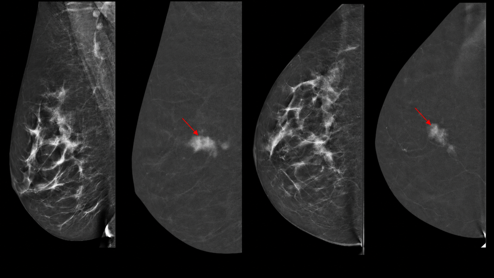
- Contrast-enhanced mammography (CEM): In addition to providing a routine mammogram, CEM highlights areas with increased blood flow, a hallmark of many cancers. Like breast MRI, CEM relies on contrast to “light up” hidden cancers, providing an important alternative for people who get anxious in the confined space of an MRI machine. Access to CEM remains limited in Los Angeles, but the technology is available through RadNet’s Breastlink—and it’s significantly less expensive than breast MRI.
How Dense Breast Tissue Impacts Screening
Breast tissue is made up of glandular tissue, fibrous (connective) tissue and fat. Dense breast tissue has larger amounts of glandular and fibrous tissue and lower amounts of fatty tissue.
About half of women aged 40 and older have dense breasts. Women with extremely dense breasts face a two- to three-fold increased risk of developing breast cancer compared to those with almost entirely fatty tissue. A mammogram is the only way to find out if you have dense breasts.
“Dense breast tissue not only increases breast cancer risk, but it also makes cancers more difficult to detect,” said Cullinane. “Both dense breast tissue and abnormal changes in the breast appear as solid white on a mammogram—unlike fatty tissue, which is black.”
While there is no single screening guideline for women with dense breasts, many clinicians recommend annual 3D mammography beginning at age 40, with supplemental imaging for women who fall into a higher-risk category.
{{cta-block}}
How AI Is Changing Breast Imaging
Improved imaging has saved lives by detecting cancers earlier than ever before, but it has also increased the likelihood that women will be called back for additional imaging after routine screening.
“In my experience in over a decade of screening, 30% to 50% of women will receive at least one call back, which requires additional imaging and a possible biopsy,” said Cullinane. “And most ultimately learn they do not have cancer.”
As imaging technology becomes even more sensitive, clinicians are continuing to try to strike a balance between detecting cancer early and minimizing unnecessary biopsies, procedures and anxiety.
Here’s the good news: AI holds tremendous promise for identifying cancers that radiologists might otherwise miss and helping distinguish which findings need further evaluation. It’s also helping clinicians better understand each woman’s individual risk.
Recently, the National Comprehensive Cancer Network published its 2026 Clinical Practice Guidelines for breast cancer screening to include an AI-based risk-assessment tool that analyzes mammograms to calculate a woman’s five-year risk of developing breast cancer beginning at age 35.
“When these tools become widely available, we’ll be better equipped to help women make informed decisions, not just about screening, but also about the best way to treat cancers should they develop,” said Jochelson.
Questions to Ask Your Doctor About Breast Cancer Screening
Your doctor can help you make sense of different screening recommendations and determine which approach makes sense for you. Consider asking:
- When should I start getting mammograms?
- How often should I be screened?
- Do I have dense breast tissue?
- Does my family history change my screening schedule?
- Should I consider genetic counseling or testing?
- Do I need ultrasound or MRI in addition to mammography?
- Could my screening plan change over time?
Build a Screening Plan Around Your Risk
Guidelines differ on timing, but if your risk of developing breast cancer is above 20%, your doctor likely will recommend a more intensive screening strategy, including:
- Earlier screening based on family history and other personal risk factors
- More frequent or advanced imaging, such as annual mammograms plus breast MRI
- Genetic counseling or genetic testing for personalized prevention strategies
- Preventive medications, such as tamoxifen, raloxifene or aromatase inhibitors for women at highest risk
- Certain breast screening findings or elevated risk scores may also prompt a referral to a breast surgeon to discuss risk-reducing surgery, preventive medications and a personalized surveillance plan.
“In the future, genetic testing, coupled with radiographic images and AI, may become the norm for every woman to carefully review all of their risk factors and then receive tailored recommendations for screening and possibly prevention strategies,” said Jochelson.
Frequently Asked Questions
Can your breast cancer screening plan change over time?
Yes. Screening recommendations aren’t fixed. Age, changes in breast density, new family history, updated genetic test results or a new personal health diagnosis can all shift screening recommendations.
Does a normal mammogram mean you don’t need any other breast imaging?
Not necessarily. Whether you need supplemental imaging—such as ultrasound, MRI or contrast-enhanced mammography—depends on your individual risk factors, breast density and any findings from your mammogram, not just whether the mammogram itself came back normal.
Does having dense breasts mean you will develop breast cancer?
No. Dense breast tissue increases your relative risk of developing breast cancer and can make cancers harder to spot on a mammogram, but it does not mean you will develop the disease.
Do breast implants change how mammograms are performed?
Patients with breast implants should still get mammograms, but it’s important to notify the imaging center ahead of time. Additional views or specialized techniques are often needed to get a complete, accurate picture of the breast tissue around the implant. Women who have had a bilateral mastectomy with reconstructive implants don’t need mammograms, unless their physician said otherwise.
What information should I share with my doctor when discussing breast cancer screening?
Come prepared to discuss your family history of breast and ovarian cancer; any prior breast findings, biopsies or procedures, or genetic testing you’ve had (or are considering); your breast density, if known; and any other personal risk factors—such as reproductive history or prior chest radiation.