
Anatomy of the Eye
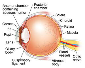
The structures of the eye include the cornea, iris, pupil, macula, retina, and the optic nerve.
On this page
- Anterior chamber. The front section of the eye's interior where aqueous humor flows in and out, providing nourishment to the eye.
- Anterior chamber. The front section of the eye's interior where aqueous humor flows in and out, providing nourishment to the eye.
- Aqueous humor. The clear watery fluid in the front part of the eye known as the anterior chamber.
- Blood vessels. Tubes (arteries and veins) that carry blood to and from the eye.
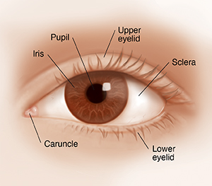
- Caruncle. A small, red structure in the corner of the eye closest to the nose that contains modified sebaceous and sweat glands.
- Choroid. The thin layer of blood vessels that lies between the retina and the sclera and supplies blood to the outer portion of the retina.
- Ciliary body. The part of the eye that makes aqueous humor.
- Cornea. The clear, dome-shaped surface that covers and protects the front of the eye.
- Iris. The colored part of the eye that helps control the amount of light entering the eye.
- Lens (crystalline lens). The transparent structure inside the eye that focuses light rays onto the retina, much like the lens in a camera.
- Lower eyelid. Skin that covers the lower part of the eye, including the cornea, when closed.
- Macula. The central portion of the retina that allows us to see fine details.
- Optic nerve. A bundle of nerve fibers that connect the retina with the brain. The optic nerve carries signals of light, dark, and colors to a part of the brain called the visual cortex, which assembles the signals into images and results in vision.
- Posterior chamber. The very narrow part of the eye behind the iris and in front of the lens
- Pupil. The opening in the center of the iris through which light passes to the back of the eye.
- Retina. The light-sensitive nerve layer that lines the inside of the back of the eye. The retina senses light and creates impulses that are sent through the optic nerve to the brain.
- Sclera. The white visible portion of the eye. The muscles that move the eye are attached to the sclera.
- Suspensory ligament of lens. A series of fibers that connects the ciliary body with the lens, holding it in place.
- Upper eyelid. Skin that covers the upper part of the eye, including the cornea, when closed.
- Vitreous body. A clear, jelly-like substance that fills the middle of the eye between the lens and the retina.
Care and Services
Explore the care options and services we offer at Cedars-Sinai.
Were always here to help.
Get in touch with us by calling 1‑800-CEDARS-1 (1‑800-233-2771) , Monday‑Friday, 8 am‑5 pm PT or feel free to send us a message.