Pathologists Go Beyond the Glass Slide
Date
February 24, 2021
Credits

Date
February 24, 2021
Credits
Medical providers featured in this article
In Brief
{{cta-block}}
For more than a century, upgraded versions of the classic light microscope have remained among the most important tools in a pathologist’s arsenal. Before the coronavirus pandemic, pathology faculty and residents routinely scrutinized the same sample together on a glass slide through a multi-headed microscope, their faces just inches apart.
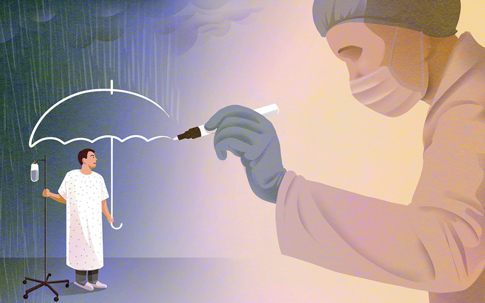
Infection-control measures to stop the spread of COVID-19 forced pathologists to step at least six feet away from each other—and to leap into the future.
“With a digital slide, we can enable residents, attending physicians and others to look at the specimen in different locations without driving the glass slide all over town.”
Because of the pandemic, the Food and Drug Administration relaxed its requirements for digital pathology devices and allowed scanned slides to be used to make primary diagnoses rather than requiring a glass slide to be seen in person.
The scans are rich reproductions of tissue samples, allowing doctors to zoom in on areas of interest just as they would with a microscope. This allows for accurate measurements and digital annotations.
“With COVID-19, the terrain is changing somewhat for digital pathology,” says David Frishberg, MD, interim chair of Pathology and Laboratory Medicine. “With a digital slide, we can enable residents, attending physicians and others to look at the specimen in different locations without driving the glass slide all over town.”
Cedars-Sinai typically produces up to 1,000 new tissue glass slides per day, and these slides will continue to be a necessary starting point for the foreseeable future, as samples will still be dissected, processed and treated with special stains before they are digitally scanned.
Digital slides are also proving useful in medical huddles, such as tumor boards, where multiple specialists examine a digital specimen in real time rather than viewing static photos. They could also be useful for new cases, allowing several doctors to review a sample quickly to make a diagnosis. In the future, digital slides may be used in conjunction with artificial intelligence algorithms, potentially leading to quicker and more accurate diagnoses, according to Frishberg.