Research Areas
Evolution During Chronic Cystic Fibrosis Infection and Antibiotic Resistance
The Jorth Laboratory is interested in how microbes evolve during infection to escape the immune response and resist antibiotic treatment. Individuals with cystic fibrosis (CF) suffer from lifelong chronic bacterial lung infections, primarily caused by Pseudomonas aeruginosa (P. aeruginosa). The Jorth Lab uses genomic approaches to:
- Identify mutations that arise in bacteria collected from CF lung infections
- Discover mutations that arise in laboratory-evolved antibiotic resistant bacteria
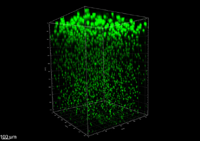
P. aeruginosa biofilm aggregates grown in an agar block biofilm assay. Bacteria (green) were stained with HCR nucleic acid probes and imaged by confocal microscopy after 16 hours growth. Biofilms promote bacterial tolerance to antibiotic treatment and play an important role during chronic CF infections. Biofilm aggregates growing near oxygen are larger at the top of the agar block than aggregates growing with less oxygen near the bottom of the agar block.
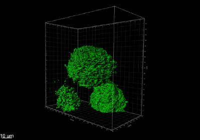
P. aeruginosa biofilm aggregates grown in an agar block biofilm assay. Bacteria (green) were stained with HCR nucleic acid probes and imaged by confocal microscopy after 16 hours growth. Aggregate surfaces were reconstructed from HCR nucleic acid staining.
Virulence and Host-Pathogen Interactions
The Jorth Lab recently discovered a set of P. aeruginosa virulence-enhancing resistance mutations (VERMs) that confer aztreonam antibiotic resistance and increase P. aeruginosa virulence, even in the absence of antibiotics. Lab members are working to understand the molecular mechanisms by which these VERMs simultaneously make this bacterium more resistant to treatment and lethal to the host.
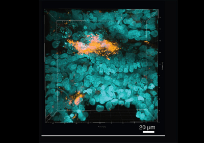
Bacterial biofilm in CF lung tissue. Image captured by confocal microscopy after tissue clearing, immunofluorescence staining for P. aeruginosa (orange), and DAPI staining of host nuclei (blue).
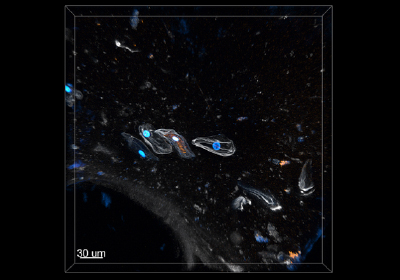
Bacteria inside human cells CF sputum. Image captured by confocal microscopy after tissue clearing, hybridization chain reaction (HCR) nucleic acid staining for P. aeruginosa (orange), DAPI staining of host nuclei (blue), lectin staining of host mucin (white).
Microbiome
The human body is essentially a culture vessel for countless microbes that colonize our bodies. In addition to causing disease, these organisms play essential roles in immune system development and help protect us from infection by invading pathogens. The Jorth Laboratory is interested in how the microbiome changes both compositionally and functionally during different disease states and treatments. We have experience studying the lung and sputum microbiota in people with CF, as well as the periodontal microbiota in people with periodontal disease.
Contact the Jorth Lab
127 S. San Vicente Blvd.
Advanced Health Sciences Pavilion, Room 9600
Los Angeles, CA 90048