Research Areas
Development of novel MR techniques for neurovascular imaging
Neurovascular disease is one of the major causes of morbidity and mortality worldwide and is the number one cause of adult disability. Dr. Fan’s lab has been focused on the development of 3D multi-contrast-weighting MR techniques for better diagnosis or risk stratification of various neurovascular diseases. Representative achievements are:
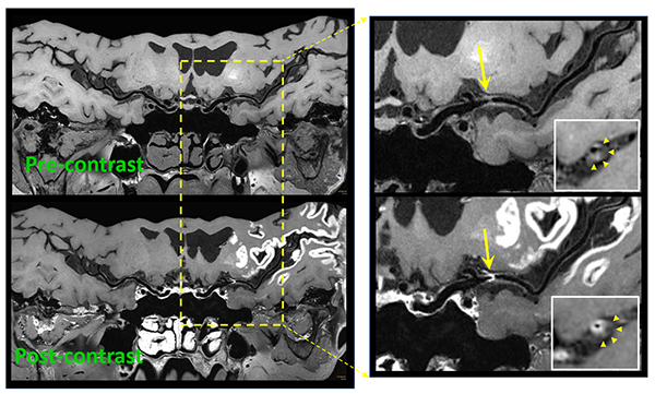
Figure: Reformatted intracranial vessel wall images acquired with the whole-brain MR vessel wall imaging technique. Upper row: images without gadolinium-based contrast depict a large-burden atherosclerotic plaque at the middle cerebral artery. Lower row: images with contrast injection depict strong plaque enhancement suggestive of intraplaque active inflammation.
We developed a multi-contrast atherosclerosis characterization (MATCH) technique (United States Patent US9554727B2) that represents the first 3D method allowing for comprehensive carotid atherosclerotic plaque composition assessment in 5 minutes. This dramatically mitigates the limitations (such as inter-scan misalignment, suboptimal tissue contrast, lengthy exam duration) associated with conventional MR carotid plaque imaging techniques, thus potentially fostering clinical adoption of MR in guiding management of carotid atherosclerotic disease. Dr. Fan has served as a co-author in the White Paper on carotid vessel wall imaging from the American Society of Neuroradiology.
We recently developed a cerebrospinal fluid-suppressed whole-brain vessel wall imaging technique that allows for non-invasive characterization of intracranial vessel wall with superior spatial resolution and image contrast. Dr. Fan is one of multiple Principal Investigators leading a multi-center registry (“WISP”, ClinicalTrials.gov NCT02923752) to evaluate the technique’s clinical utility in stroke etiology determination.
Dr. Fan was awarded by NIH/NHLBI in 2019 an R01 grant to develop a longitudinal and quantitative MR plaque imaging technique for treatment response evaluation in symptomatic intracranial atherosclerosis.
- Fan Z, Yu W, Xie Y, Dong L, Yang L, Wang Z, Conte A.H., Bi X, An J, Zhang T, Laub G, Shah P.K., Zhang Z, Li D. Multi-contrast atherosclerosis characterization (MATCH) of carotid plaque with a single 5-min scan: technical development and clinical feasibility. Journal of Cardiovascular Magnetic Resonance 2014;16:53.
- Fan Z, Yang Q, Deng Z, Li Y, Bi X, Song S, Li D. Whole-brain intracranial vessel wall imaging at 3 Tesla using cerebrospinal fluid-attenuated T1-weighted 3D turbo spin echo. Magn Reson Med. 2017 Mar;77(3):1142-1150.
- Wu F, Song H, Ma Q, Xiao J, Jiang T, Huang X, Bi X, Guo X, Li D, Yang Q, Ji X, Fan Z. Hyperintense Plaque on Intracranial Vessel Wall Magnetic Resonance Imaging as a Predictor of Artery-to-Artery Embolic Infarction. Stroke 2018;49:905-911.
- Saba L, Yuan C, Hatsukami TS, Balu N, Qiao Y, DeMarco JK, Saam T, Moody AR, Li D, Matouk CC, Johnson MH, Jager HR, Mossa-Basha M, Kooi ME, Fan Z, Saloner D, Wintermark M, Mikulis DJ, Wasserman BA. Carotid Artery Wall Imaging: Perspective and Guidelines from the ASNR Vessel Wall Imaging Study Group and Expert Consensus Recommendations of the American Society of Neuroradiology. American Journal of Neuroradiology 2018;39:E9-E31.
- Shi F, Yang Q, Guo X, Qureshi T, Tian Z, Miao H, Dey D, Li D, Fan Z. Intracranial Vessel Wall Segmentation Using Convolutional Neural Networks. IEEE Transactions on Biomedical Engineering 2019;66:2840-2847.
Development of novel MR techniques for guiding radiation therapy of cancer
Over the past few years, there is a keen interest in the integration of MR alone into radiation treatment planning and even the therapy workflow, i.e. MR-guided radiotherapy (MRgRT), to leverage its superior soft-tissue contrast. The abdomen, however, represents a challenging treatment site for pursuing these applications, in part due to respiratory motion and the presence of many organs-at-risk (OARs). Dr. Fan’s lab has developed several techniques related to image acquisition and post-processing of 4D-MR that can reliably depict abdominal organs and tumors and assess their respiratory motion trajectories. Representative achievements are:
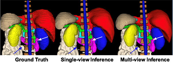
Figure: Segmentation results on a random test case with manual label, single-view (transversal) inference of DenseUnet, and multi-view inference with majority voting: liver(red), spleen(gray), pancreas(green), right kidney(blue), left kidney(yellow), stomach(cyan), duodenum(purple), small intestine(white), spinal cord (blue) and vertebral bodies (dark brown). The multi-view inference correctly segmented the small intestine that is missed in single-view inference, as shown by the blue arrow. Besides, it produced a more accurate boundary of the pancreas and duodenum, as shown by the white arrow.
Our team developed a 4D-MR technique based on 3D continuous radial-sampling spoiled gradient-echo for data acquisition and self-gating for respiratory motion detection. Data were retrospectively sorted into different respiratory phases, and each phase was reconstructed by means of self-calibrating CG-SENSE that can well handle undersampling problems. This technique eliminates the need for external motion surrogate set up, allows for the exclusion of irregular breathing cycles after scanning, and facilitates the reconstruction of an averaged phase-resolved isotropic-spatial-resolution volumetric image series with consistent image quality throughout all respiratory phases and subjects. A post-processing method, named iterative motion correction and average (United States Patent US10605880B2), was later developed to significantly improve 4D-MR image quality.
We are currently working on automated organ segmentation on MR images. Accurate and efficient delineation of the target and OARs is highly desirable for RTP, particularly for online adaptive MRgRT. However, manual delineation is still a common practice and a well-known time-consuming and interobserver variation-prone process. Deep learning has recently demonstrated great promise in tackling complex multi-organ segmentation problems in MR images. We have recently proposed a 2D DenseUnet approach to perform automated segmentation on 3D T1-weighted abdominal images. A multi-view approach by performing parallel 2D-based segmentation in the axial, coronal and sagittal views was proposed to considerably improve segmentation accuracy.
Dr. Fan was awarded by NIH/NCI in 2019 an R21 grant to develop a 4D-MR technique for monitoring pancreatic tumor infiltrating blood vessels and tumor response to chemoradiation therapy. An R01 project will also be funded by NIH/NIBIB in Summer 2020, aiming to develop a multi-task MR imaging technique for abdominal radiotherapy planning.
- Yang W, Fan Z, Tuli R, Deng Z, Pang J, Wachsman A, Reznik R, Sandler H, Li D, Fraass BA. Four-Dimensional Magnetic Resonance Imaging With 3-Dimensional Radial Sampling and Self-Gating-Based K-Space Sorting: Early Clinical Experience on Pancreatic Cancer Patients. International Journal of Radiation Oncology, Biology, Physics. 2015;93:1136-1143.
- Deng Z, Pang J, Yang W, Yue Y, Sharif B, Tuli R, Li D, Fraass B, Fan Z. Four-dimensional MRI using three-dimensional radial sampling with respiratory self-gating to characterize temporal phase-resolved respiratory motion in the abdomen. Magnetic Resonance in Medicine 2016;75:1574-1585.
- Deng Z, Pang J, Lao Y, Bi X, Wang G, Chen Y, Fenchel M, Tuli R, Li D, Yang W, Fan Z. A post-processing method based on interphase motion correction and averaging to improve image quality of 4D magnetic resonance imaging: a clinical feasibility study. British Journal of Radiology 2019;92:20180424.
- Chen Y, Ruan D, Xiao J, Wang L, Sun B, Saouaf R, Yang W, Li D, Fan Z. Fully automated multi-organ segmentation in abdominal magnetic resonance imaging with deep neural networks. arXiv:1912.11000.
Development of novel MR techniques for diagnosis of cardiovascular diseases
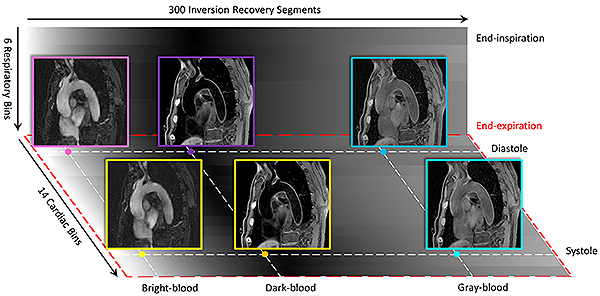
Figure: Illustration of different time dimensions for MT-MACS, namely, cardiac pulsation, respiration and T2-prepared inversion recovery dimensions. For phase-resolved imaging, MT-MACS divides cardiac motion and respiratory movement into 14 and 6 bins, respectively. Within each inversion recovery period, MT-MACS contains 300 readout segments which can generate 300 various contrast weightings. 3 typical contrast weightings (i.e. bright-blood, dark-blood, and gray-blood) are picked out for multi-contrast assessment.
Cardiovascular diseases, as a group of disorders of the heart and blood vessels, are the number 1 cause of death globally. Early diagnosis of cardiovascular diseases is of paramount importance. Dr. Fan’s lab has invested a great deal of effort in the development of non-contrast-enhanced MR techniques for accurate diagnosis of various cardiovascular diseases, such as cardiac abnormalities, peripheral arterial disease, deep vein thrombosis, and thoracic aortic disease.
We recently develop an MR MultiTasking (MT) based multi-dimensional assessment of cardiovascular system (MACS) with ECG- and navigator-free data acquisition to enable a comprehensive evaluation of thoracic aorta. The MT-MACS method adopted a low-rank tensor imaging model with a cardiac time dimension for phase-resolved cine imaging and a T1 recovery dimension for multi-contrast assessment. MT-MACS images reconstructed from acquisitions as short as 6 minutes demonstrated good or excellent image quality for bright-blood, dark-blood, and gray-blood contrast weightings, respectively. Analysis showed good agreement in the lumen and wall area and aortic strain between MT-MACS and conventional 2D sequences.
- Xie G, Bi X, Liu J, Yang Q, Natsuaki Y, Conte A.H., Liu X, Li K, Li D, Fan Z. Three-dimensional coronary dark-blood interleaved with gray-blood (cDIG) magnetic resonance imaging at 3 tesla. Magnetic Resonance in Medicine 2016;75:997-1007. PMID: 25858528.
- Xie G, Chen H, He X, Liang J, Deng W, He Z, Ye Y, Yang Q, Bi X, Liu X, Li D, Fan Z. Black-blood thrombus imaging (BTI): a contrast-free cardiovascular magnetic resonance approach for the diagnosis of non-acute deep vein thrombosis. Journal of Cardiovascular Magnetic Resonance 2017;19:4. PMID: 28095878.
- Zhang X, Xie G, Zhu Y, Wei Z, Su S, Shi C, Yan F, Liu X, Qiu B, Fan Z. 3D self-gated cardiac cine imaging at 3 Tesla using stack-of-stars bSSFP with tiny golden angles and compressed sensing. Magnetic Resonance in Medicine. 2019;81:3234-3244. PMID: 30474151.
- Hu Z, Christodoulou A, Wang N, Shaw J, Song S, Maya M, Ishimori, M, Forbess L, Xiao J, Bi X, Han F, Li D, Fan Z. Magnetic resonance multitasking for multidimensional assessment of cardiovascular system: Development and feasibility study on the thoracic aorta. Magnetic Resonance in Medicine 2020 (In press).
Contact the Fan Lab
Cedars-Sinai Biomedical Imaging Research Institute
8700 Beverly Blvd.
Pacific Theatres Building, Suite 400
Los Angeles, CA 90048